
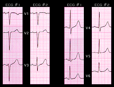
Variations in the relatioship of the chest leads to the heart itself, caused most often by changes in the placement of the chest leads or, less often by changes in the patient's position, can impact significantly on the QRS waveform. This is most important in the transitional leads (leads V3 and V4)since these are the leads in which the QRS complex changes from one in which the S wave is of greater amplitude than the R wave, to one in which the R wave has a larger amplitude than the S wave.
The two ECGs shown here were recorded one week apart on the same 82 year old patient with no known heart disease. Focus your attention on lead V3. In ECG #1, V3 shows a small R wave followed be a large S wave, whereas In ECG #2, the R wave is of greater amplitude than the S wave. The other leads differ slightly in the amplitude of the QRS complexes but not in their morphology. The change in V3 is caused by a change in the position of the V3 lead rather than by a change in the electrical activity of the heart. Such artifactual changes may cause inappropriate ECG diagnoses if the reader is not aware of the the effect of even slight changes in the placement of the chest leads.