
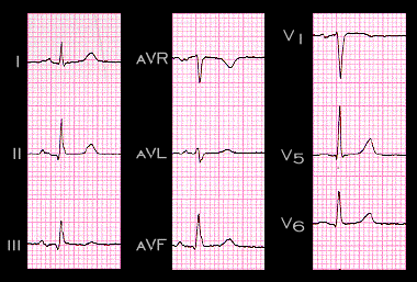
In this ECG, there is a small, narrow Q wave in leads II, III and aVF, and a small narrow R wave in lead aVL, These findings indicate that septal depolarization is more superiorly directed. The small, narrow R wave in V1 reflects the anterior orientation of septal depolarization.