
The upper tracing is from the same patient but now, the arm leads have been reversed and the chest leads have been placed on the right side of the chest. Below it is the tracing from the previous page, recorded with the leads placed in their usual positions.
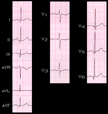
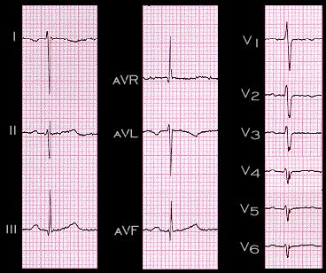
Reversing the arm leads normalizes the P waves in the limb leads. The P wave in lead I is changed from negative to positive, in lead aVL from negative to isoelectric and in lead aVR from isoelectric to negative. With the chest leads placed n the right side of the chest, the P wave becomes isoelectric in lead V1 ,slightly positive in lead V2. and slightly more positive in leads V4-6. The QRS complexes and T waves are also changed and now appear normal. Thus, in mirror image dextrocardia, the ECG abnormalities are normalized by reversing the placement of the arm leads and by placing the chest leads on the right side of the chest.