
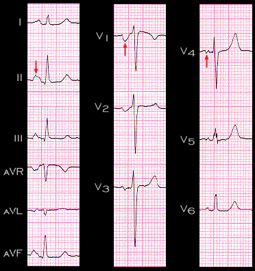
A second common P wave abnormality is often seen in patients with mitral valve disease and has been labeled "P mitrale". This ECG is from a patient with mitral stenosis and insufficiency. The P wave duration is 130 ms. The prolonged duration, seen best in leads II and V1, is primarily due to the increased duration of the second half of the P wave, i.e. that portion associated with left atrial depolarization which, in lead V1, is the negative component. Note that this P wave component is increased in both duration and amplitude Note also that in lead II, there is a notch that separates the right and left atrial components. In this patient the notch is also prominent in leads V4 and V5, giving the P wave an M-shaped appearance.