Home
Understanding the Electrocardiogram

Main navigation

  • Home
  • Understanding ECGs
    • Introduction
User account menu
  • Log in

Breadcrumb

  1. Home
  2. Understanding the Electrocardiogram In Health & Disease
  3. 2.0.0 The Normal Electrocardiogram
  4. 2.1.0 P Wave

2.1.17

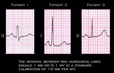
The amplitude of the normal P wave does not usually exceed 2.5mm (0.25 mV) at the standard calibration of 10 mm per mV. This figure shows the tallest P wave and the lead in which they were recorded from each of the three patients whose ECGs were shown earlier. A number of factors, such as the patient's body build, the presence of pericardial or pleural effusions or of tissue edema, and the position of the heart within the thorax will impact on the proximity of the electrodes to the heart and/or on electrical conductivity and thus on the amplitude of the P wave (as well as that of the QRS and T wave) recorded on the body surface.

Book traversal links for 2.1.17

  • 2.1.16
  • Up
  • 2.1.18

Site is under construction

Book navigation

  • Introduction to First Edition
  • 1.0.0 Generation of the ECG
  • 2.0.0 The Normal Electrocardiogram
    • 2.1.0 P Wave
      • 2.1.1
      • 2.1.2
      • 2.1.3
      • 2.1.4
      • 2.1.5
      • 2.1.6
      • 2.1.7
      • 2.1.8
      • 2.1.9
      • 2.1.10
      • 2.1.11
      • 2.1.12
      • 2.1.13
      • 2.1.14
      • 2.1.15
      • 2.1.16
      • 2.1.17
      • 2.1.18
      • 2.1.19
      • 2.1.20
      • 2.1.21
      • 2.1.22
      • 2.1.23
      • 2.1.24
      • 2.1.25
      • 2.1.26
      • 2.1.27
      • 2.1.28
      • 2.1.29
      • 2.1.30
      • 2.1.31
      • 2.1.32
      • 2.1.33
    • 2.2.0 PR Interval
    • 2.3.0 QRS Complex
    • 2.4.0 ST segment
    • 2.5.0 T wave
    • 2.6.0 QT Interval
    • 2.7.0 U Wave
  • 3.0.0 Inter and intra-ventricular Conduction Disturbances
  • 4.0.0 Ventricular Hypertrophy
  • 5.0.0 ELECTROLYTE ABNORMALITIES, DRUG EFFECTS AND THE LONG QT SYNDROMES
  • 6.0.0 Ischemia and Infarction - Introduction (frame i and ii)
  • 7.0.0 Tachycardias- Introduction
  • 8.0.0 The Bradycardias frame i-introduction
  • 9.0.0 The ECG of Heart Murmurs-introduction
  • 10.0.0 The Electrocardiogram in the Emergency Department-Introduction
Powered by Drupal