
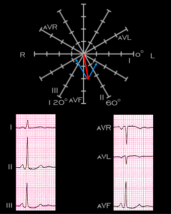
In contrast to the ECG shown on pages 2.1.1 - 2.1.6, in which the P wave was flat, or isoelectric, in lead aVL, the P wave in this tracing is negative in lead aVl. This is shown by the projection of the P wave position to the perpendicular of aVL. The P wave remains negative in lead aVR.