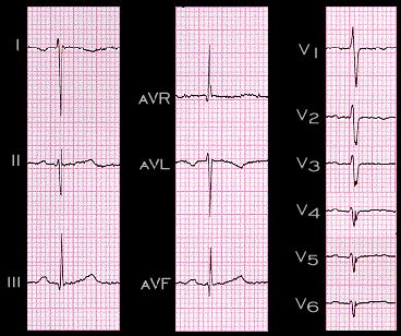
The ECG shown here is from a patient with mirror image dextrocardia. The leads are placed in their normal positions. Note that the P wave is inverted in lead I and aVL and is isoelectric in lead aVR, as in the patient with the reversed arm leads. In the chest leads, the P waves still appear to be within normal limits. The more obvious abnormalities are in the QRS complex and these will be discussed later.