
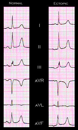
An ectopic atrial pacemaker is probably the cause of the different P wave configurations in the 2 ECGs from the same patient shown here. The P wave in the tracing on the left is normal. Its frontal plane axis of approximately 60 degrees reflects its origin within the sinus node. The P wave in the tracing on the right is abnormal. It is inverted in leads II,III and aVF and isoelectric in aVR. Its frontal plane axis is -38 degrees indicating a pacemaker located outside of the sinus node. The leftward and superior direction of its spatial vector suggests a location somewhere in the lower portion of the right atrium.