
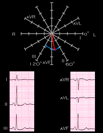
Focus your attention on the P wave in this electrocardiagram. It has an amplitude of about +2 mm in each of leads II, III and aVF and less than +1 mm in lead I. As shown in the figure, its electrical axis in the frontal plane is +88 degrees and its spatial vector is directed inferiorly.