
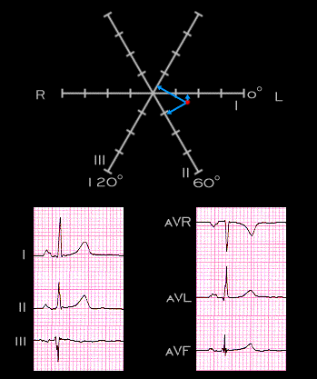
This ECG is from another patient. This P wave is also of sinus origin but its electical axis in the frontal plane is more horizontal (+10 degrees ). It is slightly more positive in lead I than in lead II and is slightly negative in lead III. This is in marked contrast to both prior tracings in which the P wave was positive in lead III.