
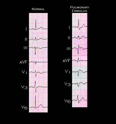
Selected leads from the two tracings are shown here. The changes associated with the pulmonary embolus shown here are quite classical. Frequently, differentiating these changes from those associated with an acute inferior wall myocardial infarction is difficult. It is often accomplished on the basis of the presenting symptoms and the absence of Q waves in lead II in patients with an acute pulmonary embolus.