
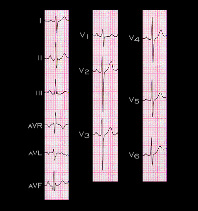
The ECG shown here is from a 35 year old male with COPD of moderate severity. It shows many features that are similar, albeit less pronounced, than those that were present in the previous patient. The axis is slightly less rightward (+125 degrees) and the P wave changes in the limb leads Leads are less dramatic. There is a persistent S wave in leads V4, V5 and V6, but the R wave is taller and the R to S ratio in V6 is greater than 1. In general, the more severe the chronic lung disease, the more marked the electrocardiographic changes.