Home
Understanding the Electrocardiogram

Main navigation

  • Home
  • Understanding ECGs
    • Introduction
User account menu
  • Log in

Breadcrumb

  1. Home
  2. Understanding the Electrocardiogram In Health & Disease
  3. 1.0.0 Generation of the ECG
  4. 1.9.0 ECG Leads and Placement

1.9.18

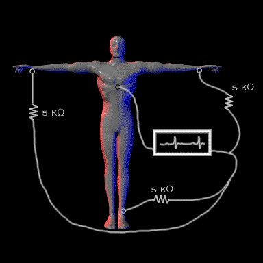
The concept of the unipolar leads was originally developed by Dr. Frank Wilson, a student of Sir Thomas Lewis. It was Dr. Wilson who brought the electrocardiogram to the United States from England. Dr. Wilson connected a resistor of 5,000 ohms to each of the limb leads in order to minimize effects of skin resistance and then combined the leads to form the indifferent reference electrode,. This is referred to as the "Central Terminal." He then  positioned an exploring electrode at various locations on the chest wall to record electrical activity that was closer to the heart than that recorded by the limb leads.

You should recognize that the chest leads are not truly unipoplar because the reference, or indifferent lead is comprised of the combined limb leads rather than ground. Thus, although the term "unipolar" is a misnomer, it has become part of the ECG vernacular and is likely to remain so.

Book traversal links for 1.9.18

  • 1.9.17
  • Up
  • 1.9.19

Site is under construction

Book navigation

  • Introduction to First Edition
  • 1.0.0 Generation of the ECG
    • 1.1.0 Basic Physiology
    • 1.2.0 SA Node & Pacemakers
    • 1.3.0 Atrial Depolarization
    • 1.4.0 Atrio Ventricular Conduction
    • 1.5.0 Ventricular Depolarization
    • 1.6.0 Repolarization
    • 1.7.0 Events at the Cellular Level
    • 1.8.0 Electrodes
    • 1.9.0 ECG Leads and Placement
      • 1.9.1
      • 1.9.2
      • 1.9.3
      • 1.9.4
      • 1.9.5
      • 1.9.6
      • 1.9.7
      • 1.9.8
      • 1.9.9
      • 1.9.10
      • 1.9.11
      • 1.9.12
      • 1.9.13
      • 1.9.14
      • 1.9.15
      • 1.9.16
      • 1.9.17
      • 1.9.18
      • 1.9.19
      • 1.9.20
      • 1.9.21
      • 1.9.22
      • 1.9.23
      • 1.9.24
      • 1.9.25
      • 1.9.26
      • 1.9.27
      • 1.9.28
      • 1.9.29
  • 2.0.0 The Normal Electrocardiogram
  • 3.0.0 Inter and intra-ventricular Conduction Disturbances
  • 4.0.0 Ventricular Hypertrophy
  • 5.0.0 ELECTROLYTE ABNORMALITIES, DRUG EFFECTS AND THE LONG QT SYNDROMES
  • 6.0.0 Ischemia and Infarction - Introduction (frame i and ii)
  • 7.0.0 Tachycardias- Introduction
  • 8.0.0 The Bradycardias frame i-introduction
  • 9.0.0 The ECG of Heart Murmurs-introduction
  • 10.0.0 The Electrocardiogram in the Emergency Department-Introduction
Powered by Drupal