Home
Understanding the Electrocardiogram

Main navigation

  • Home
  • Understanding ECGs
    • Introduction
User account menu
  • Log in

Breadcrumb

  1. Home
  2. Understanding the Electrocardiogram In Health & Disease
  3. 1.0.0 Generation of the ECG
  4. 1.9.0 ECG Leads and Placement

1.9.29

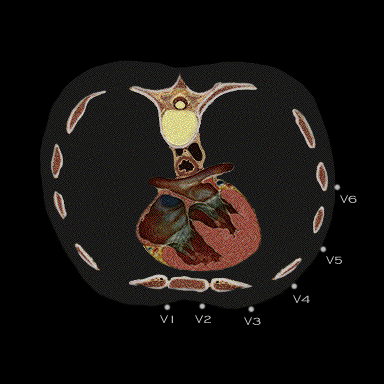
As demonstrated in this figure, leads V1 and V2 are in closest proximity to the anterior cardiac chambers and are most likely to demonstrate local electrical events. Leads V3 through V6 become progressively further distanced from the heart itself. Leads V3 and V4 are located approximately over the apex of the heart and leads V5 and V6, which are more or less in closer proximity to the left ventricular lateral wall, are least likely to demonstrate local electrical events.

Book traversal links for 1.9.29

  • 1.9.28
  • Up
  • 2.0.0 The Normal Electrocardiogram

Site is under construction

Book navigation

  • Introduction to First Edition
  • 1.0.0 Generation of the ECG
    • 1.1.0 Basic Physiology
    • 1.2.0 SA Node & Pacemakers
    • 1.3.0 Atrial Depolarization
    • 1.4.0 Atrio Ventricular Conduction
    • 1.5.0 Ventricular Depolarization
    • 1.6.0 Repolarization
    • 1.7.0 Events at the Cellular Level
    • 1.8.0 Electrodes
    • 1.9.0 ECG Leads and Placement
      • 1.9.1
      • 1.9.2
      • 1.9.3
      • 1.9.4
      • 1.9.5
      • 1.9.6
      • 1.9.7
      • 1.9.8
      • 1.9.9
      • 1.9.10
      • 1.9.11
      • 1.9.12
      • 1.9.13
      • 1.9.14
      • 1.9.15
      • 1.9.16
      • 1.9.17
      • 1.9.18
      • 1.9.19
      • 1.9.20
      • 1.9.21
      • 1.9.22
      • 1.9.23
      • 1.9.24
      • 1.9.25
      • 1.9.26
      • 1.9.27
      • 1.9.28
      • 1.9.29
  • 2.0.0 The Normal Electrocardiogram
  • 3.0.0 Inter and intra-ventricular Conduction Disturbances
  • 4.0.0 Ventricular Hypertrophy
  • 5.0.0 ELECTROLYTE ABNORMALITIES, DRUG EFFECTS AND THE LONG QT SYNDROMES
  • 6.0.0 Ischemia and Infarction - Introduction (frame i and ii)
  • 7.0.0 Tachycardias- Introduction
  • 8.0.0 The Bradycardias frame i-introduction
  • 9.0.0 The ECG of Heart Murmurs-introduction
  • 10.0.0 The Electrocardiogram in the Emergency Department-Introduction
Powered by Drupal