Home
Understanding the Electrocardiogram

Main navigation

  • Home
  • Understanding ECGs
    • Introduction
User account menu
  • Log in

Breadcrumb

  1. Home
  2. Understanding the Electrocardiogram In Health & Disease
  3. 1.0.0 Generation of the ECG
  4. 1.9.0 ECG Leads and Placement

1.9.25

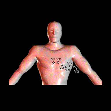
  The standard position of the exploring chest electrode in adults is as follows:

         Lead V1 is in the fourth intercostal space to the right of the sternum

         Lead V2 is in the fourth intercostal space to the left of the sternum

         Lead V3 lies between V2 and V4,

         Lead V4 is in the fifth intercostal space in the mid-clavicular line

         Lead V5 is in the fifth intercostal space in the anterior axillary line

         Lead V6 is in the fifth intercostal space in the mid-axillary line

  In women, the exploring electrode should be placed beneath the breast.

Book traversal links for 1.9.25

  • 1.9.24
  • Up
  • 1.9.26

Site is under construction

Book navigation

  • Introduction to First Edition
  • 1.0.0 Generation of the ECG
    • 1.1.0 Basic Physiology
    • 1.2.0 SA Node & Pacemakers
    • 1.3.0 Atrial Depolarization
    • 1.4.0 Atrio Ventricular Conduction
    • 1.5.0 Ventricular Depolarization
    • 1.6.0 Repolarization
    • 1.7.0 Events at the Cellular Level
    • 1.8.0 Electrodes
    • 1.9.0 ECG Leads and Placement
      • 1.9.1
      • 1.9.2
      • 1.9.3
      • 1.9.4
      • 1.9.5
      • 1.9.6
      • 1.9.7
      • 1.9.8
      • 1.9.9
      • 1.9.10
      • 1.9.11
      • 1.9.12
      • 1.9.13
      • 1.9.14
      • 1.9.15
      • 1.9.16
      • 1.9.17
      • 1.9.18
      • 1.9.19
      • 1.9.20
      • 1.9.21
      • 1.9.22
      • 1.9.23
      • 1.9.24
      • 1.9.25
      • 1.9.26
      • 1.9.27
      • 1.9.28
      • 1.9.29
  • 2.0.0 The Normal Electrocardiogram
  • 3.0.0 Inter and intra-ventricular Conduction Disturbances
  • 4.0.0 Ventricular Hypertrophy
  • 5.0.0 ELECTROLYTE ABNORMALITIES, DRUG EFFECTS AND THE LONG QT SYNDROMES
  • 6.0.0 Ischemia and Infarction - Introduction (frame i and ii)
  • 7.0.0 Tachycardias- Introduction
  • 8.0.0 The Bradycardias frame i-introduction
  • 9.0.0 The ECG of Heart Murmurs-introduction
  • 10.0.0 The Electrocardiogram in the Emergency Department-Introduction
Powered by Drupal