Home
Understanding the Electrocardiogram

Main navigation

  • Home
  • Understanding ECGs
    • Introduction
User account menu
  • Log in

Breadcrumb

  1. Home
  2. Understanding the Electrocardiogram In Health & Disease
  3. 1.0.0 Generation of the ECG
  4. 1.9.0 ECG Leads and Placement

1.9.16

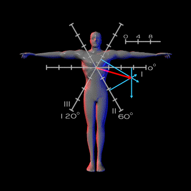
A line from the origin of the triaxial system to this point of confluence will define an angle, which in this case is +15°. This angle is defined as the main electrical vector or axis for the QRS complex.

Similar directional vectors can be created for the P wave and the T wave.

Book traversal links for 1.9.16

  • 1.9.15
  • Up
  • 1.9.17

Site is under construction

Book navigation

  • Introduction to First Edition
  • 1.0.0 Generation of the ECG
    • 1.1.0 Basic Physiology
    • 1.2.0 SA Node & Pacemakers
    • 1.3.0 Atrial Depolarization
    • 1.4.0 Atrio Ventricular Conduction
    • 1.5.0 Ventricular Depolarization
    • 1.6.0 Repolarization
    • 1.7.0 Events at the Cellular Level
    • 1.8.0 Electrodes
    • 1.9.0 ECG Leads and Placement
      • 1.9.1
      • 1.9.2
      • 1.9.3
      • 1.9.4
      • 1.9.5
      • 1.9.6
      • 1.9.7
      • 1.9.8
      • 1.9.9
      • 1.9.10
      • 1.9.11
      • 1.9.12
      • 1.9.13
      • 1.9.14
      • 1.9.15
      • 1.9.16
      • 1.9.17
      • 1.9.18
      • 1.9.19
      • 1.9.20
      • 1.9.21
      • 1.9.22
      • 1.9.23
      • 1.9.24
      • 1.9.25
      • 1.9.26
      • 1.9.27
      • 1.9.28
      • 1.9.29
  • 2.0.0 The Normal Electrocardiogram
  • 3.0.0 Inter and intra-ventricular Conduction Disturbances
  • 4.0.0 Ventricular Hypertrophy
  • 5.0.0 ELECTROLYTE ABNORMALITIES, DRUG EFFECTS AND THE LONG QT SYNDROMES
  • 6.0.0 Ischemia and Infarction - Introduction (frame i and ii)
  • 7.0.0 Tachycardias- Introduction
  • 8.0.0 The Bradycardias frame i-introduction
  • 9.0.0 The ECG of Heart Murmurs-introduction
  • 10.0.0 The Electrocardiogram in the Emergency Department-Introduction
Powered by Drupal