Home
Understanding the Electrocardiogram

Main navigation

  • Home
  • Understanding ECGs
    • Introduction
User account menu
  • Log in

Breadcrumb

  1. Home
  2. Understanding the Electrocardiogram In Health & Disease
  3. 1.0.0 Generation of the ECG

1.8.0 Electrodes

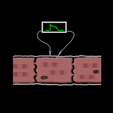
The voltage differences across the cell membrane which occur as the cell depolarizes and repolarizes can be recorded by microelectrodoes placed on opposite sides of the membrane. The generated wave form is the transmembrane action potential.

  • 1.8.1
  • 1.8.2
  • 1.8.3
  • 1.8.4
  • 1.8.5
  • 1.8.6
  • 1.8.7
  • 1.8.8
  • 1.8.9
  • 1.8.10
  • 1.8.11
  • 1.8.12
  • 1.8.13
  • 1.8.14

Book traversal links for 1.8.0 Electrodes

  • 1.7.12
  • Up
  • 1.8.1

Site is under construction

Book navigation

  • Introduction to First Edition
  • 1.0.0 Generation of the ECG
    • 1.1.0 Basic Physiology
    • 1.2.0 SA Node & Pacemakers
    • 1.3.0 Atrial Depolarization
    • 1.4.0 Atrio Ventricular Conduction
    • 1.5.0 Ventricular Depolarization
    • 1.6.0 Repolarization
    • 1.7.0 Events at the Cellular Level
    • 1.8.0 Electrodes
      • 1.8.1
      • 1.8.2
      • 1.8.3
      • 1.8.4
      • 1.8.5
      • 1.8.6
      • 1.8.7
      • 1.8.8
      • 1.8.9
      • 1.8.10
      • 1.8.11
      • 1.8.12
      • 1.8.13
      • 1.8.14
    • 1.9.0 ECG Leads and Placement
  • 2.0.0 The Normal Electrocardiogram
  • 3.0.0 Inter and intra-ventricular Conduction Disturbances
  • 4.0.0 Ventricular Hypertrophy
  • 5.0.0 ELECTROLYTE ABNORMALITIES, DRUG EFFECTS AND THE LONG QT SYNDROMES
  • 6.0.0 Ischemia and Infarction - Introduction (frame i and ii)
  • 7.0.0 Tachycardias- Introduction
  • 8.0.0 The Bradycardias frame i-introduction
  • 9.0.0 The ECG of Heart Murmurs-introduction
  • 10.0.0 The Electrocardiogram in the Emergency Department-Introduction
Powered by Drupal