
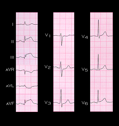
This ECG is from a 55 year old woman admitted with an acute myocarditis. The ST segment elevation in leads II, III, aVF, V5 and V6, coupled with the abnormally widened R wave and ST depression in leads V1 and V2 are all consistent with an infarction involving the inferior, posterior and lateral walls and the echocardiogram revealed wall motion abnormalities in these areas. However, her coronary angiogram was unremarkable and considered normal. Thus, as demonstrated in these last several pages, an acute myocarditis can cause ECG changes that simulate all aspects of myocardial ischemia and infarction.