Home
Understanding the Electrocardiogram

Main navigation

  • Home
  • Understanding ECGs
    • Introduction
User account menu
  • Log in

Breadcrumb

  1. Home
  2. Understanding the Electrocardiogram In Health & Disease
  3. 6.0.0 Ischemia and Infarction - Introduction (frame i and ii)
  4. 6.5.0 (frame 106) FALSE POSITIVES
  5. 6.5.20

6.5.25 (130)

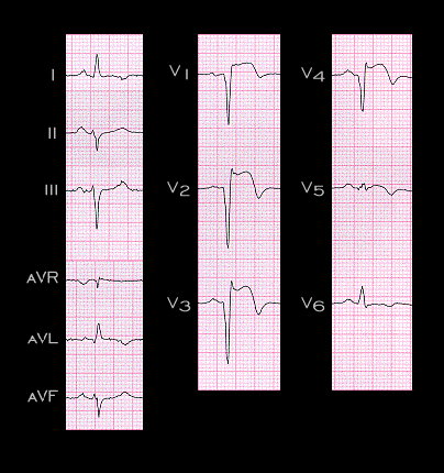
The third type of ST segment change that may simulate an acute transmural ischemia is that associated with the development of a ventricular aneurysm following an acute myocardial infarction. The ECG shown here is such an example. It is from a 65 year old patient who sustained an acute myocardial infarction three years earlier. Cardiac imaging studies revealed a well defined apical ventricular aneurysm with paradoxical wall motion. The persistence of ST segment elevation, such as that shown here, more than three weeks post infarction is suggestive of a ventricular aneurysm. It is noteworthy that within the last decade, the incidence of post infarction ventricular aneurysms has decreased significantly. This can be attributed to the application of coronary revascularization therapies within hours of the onset of an acute coronary event.

Book traversal links for 6.5.25 (130)

  • 6.5.24 (new graphic)
  • Up
  • 6.5.26 (131)

Site is under construction

Book navigation

  • Introduction to First Edition
  • 1.0.0 Generation of the ECG
  • 2.0.0 The Normal Electrocardiogram
  • 3.0.0 Inter and intra-ventricular Conduction Disturbances
  • 4.0.0 Ventricular Hypertrophy
  • 5.0.0 ELECTROLYTE ABNORMALITIES, DRUG EFFECTS AND THE LONG QT SYNDROMES
  • 6.0.0 Ischemia and Infarction - Introduction (frame i and ii)
    • 6.1.0 ST Segment (frame1)
    • 6.2.0 Q WAVES (frame 53)
    • 6.3.0 T WAVES (frame 75)
    • 6.4.0 (Bundle Branch Blocks and othe IVCDs--frame 87)
    • 6.5.0 (frame 106) FALSE POSITIVES
      • 6.5.1 (107)
      • 6.5.2 (108)
      • 6.5.3 (109)
      • 6.5.4(frame 110)
      • 6.5.5 (111)
      • 6.5.6 (112)
      • 6.5.7 (113)
      • 6.5.8 (114)
      • 6.5.9 (115)
      • 6.5.10 (116)
      • 6.5.20
        • 6.5.21 (128)
        • 6.5.22 (127)
        • 6.5.23 (frame 129)
        • 6.5.24 (new graphic)
        • 6.5.25 (130)
        • 6.5.26 (131)
        • 6.5.27 (132)
        • 6.5.28 (133)
        • 6.5.29 (frame 135)
      • 6.5.30 (frame 136)
  • 7.0.0 Tachycardias- Introduction
  • 8.0.0 The Bradycardias frame i-introduction
  • 9.0.0 The ECG of Heart Murmurs-introduction
  • 10.0.0 The Electrocardiogram in the Emergency Department-Introduction
Powered by Drupal