
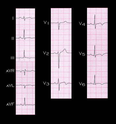
This is the ECG recorded two days later. The ST segments are no longer elevation, but now, the T waves in leads V3-V6 are inverted. These T wave changes are consistent with an acute myocarditis but they are also consistent with pericarditis and with a prior ischemic event. These T wave changes gradually resolved, as did the diffuse wall motion abnormality.