
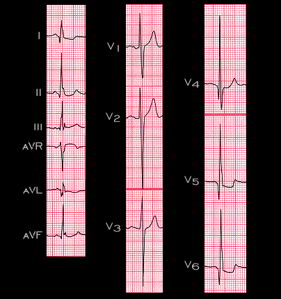
This ECG is from an 18 year of male with hypertrophic obstructive cardiomyopathy and marked asymmetric septal hypertrophy. The Q waves in leads I, aVL, V5 and V6, and the tall R wave in lead V1 reflect depolarization of the hypertrophied septum. These changes simulate and could be mistaken for those associated with an infarction of the postero-lateral wall.