Home
Understanding the Electrocardiogram

Main navigation

  • Home
  • Understanding ECGs
    • Introduction
User account menu
  • Log in

Breadcrumb

  1. Home
  2. Understanding the Electrocardiogram In Health & Disease
  3. 6.0.0 Ischemia and Infarction - Introduction (frame i and ii)
  4. 6.5.0 (frame 106) FALSE POSITIVES
  5. 6.5.10 (116)

6.5.19 (126) [ FRAME125 OMITTED]

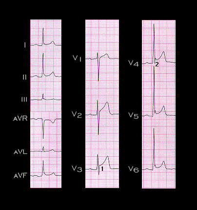
A second cause of non-ischemia related ST segment elevation is shown here. This ECG demonstrates what is referred to as “early repolarization” and is a normal variant, seen most often in younger males. It is from an asymptomatic 37 year old male with no evidence of heart disease, and was recorded as part of a routine examination. Note the following:

  1. Elevation of the ST segment at the takeoff between the end of the QRS complex and the beginning of the ST segment, seen best in this example in leads V2,and V3 (1). This is referred to as the “J point”
  2. The distinct notch at the end of the R wave, seen best in leads V4 and V5 (2)
  3. The fact that the changes are most marked in leads V2-V5, the mid-precordial leads.
  4. The magnitude of ST elevation is less than 4mm
  5. The upright and symmetrical T waves are of significantly greater (three to four times) amplitude than the ST elevation.

These are the characteristics of early repolarization

Book traversal links for 6.5.19 (126) [ FRAME125 OMITTED]

  • 6.5.18 (124)
  • Up
  • 6.5.20

Site is under construction

Book navigation

  • Introduction to First Edition
  • 1.0.0 Generation of the ECG
  • 2.0.0 The Normal Electrocardiogram
  • 3.0.0 Inter and intra-ventricular Conduction Disturbances
  • 4.0.0 Ventricular Hypertrophy
  • 5.0.0 ELECTROLYTE ABNORMALITIES, DRUG EFFECTS AND THE LONG QT SYNDROMES
  • 6.0.0 Ischemia and Infarction - Introduction (frame i and ii)
    • 6.1.0 ST Segment (frame1)
    • 6.2.0 Q WAVES (frame 53)
    • 6.3.0 T WAVES (frame 75)
    • 6.4.0 (Bundle Branch Blocks and othe IVCDs--frame 87)
    • 6.5.0 (frame 106) FALSE POSITIVES
      • 6.5.1 (107)
      • 6.5.2 (108)
      • 6.5.3 (109)
      • 6.5.4(frame 110)
      • 6.5.5 (111)
      • 6.5.6 (112)
      • 6.5.7 (113)
      • 6.5.8 (114)
      • 6.5.9 (115)
      • 6.5.10 (116)
        • 6.5.11 (frame 117)
        • 6.5.12 (118)
        • 6.5.13 (119)
        • 6.5.14 (frame 120)
        • 6.5.15 (121)
        • 6.5.16 (122)
        • 6.5.17 (123)
        • 6.5.18 (124)
        • 6.5.19 (126) [ FRAME125 OMITTED]
      • 6.5.20
      • 6.5.30 (frame 136)
  • 7.0.0 Tachycardias- Introduction
  • 8.0.0 The Bradycardias frame i-introduction
  • 9.0.0 The ECG of Heart Murmurs-introduction
  • 10.0.0 The Electrocardiogram in the Emergency Department-Introduction
Powered by Drupal