
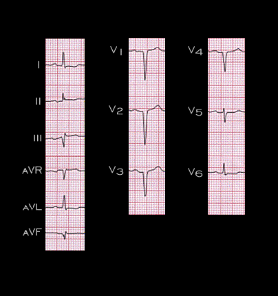
The ECG shown here is from a 48 year old woman with cardiac amyloidosis but a normal coronary angiogram. There are abnormal Q waves in III and aVF and a QS complex in leads V1-V4 simulating an anterior and possibly an inferior wall infarction. ECG abnormalities of this type are quite common in patients with cardiac amyloidosis.