
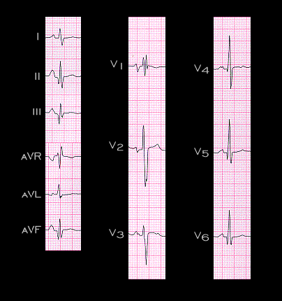
This ECG is from an 18 year old patient with Becker’s muscular dystrophy and congestive heart failure. This neuro-muscular disease, like Duchenne’s muscular dystrophy and Friedrich’s ataxia is associated with myocardial fibrosis and, like these diseases, a frequent cause of pseudo-infarction patterns on the electrocardiogram. The ECG shows incomplete right bundle block and Q waves in leads II, III, aVF, V5 and V6 that simulate the changes of an inferior wall infarction with lateral wall involvement.