
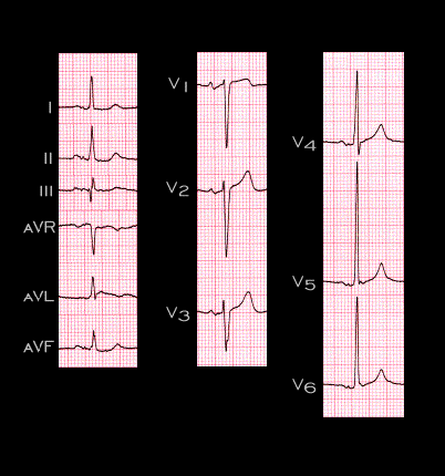
This ECG is from a 58 year old male with of both the aortic and mitral valves due to rheumatic heart disease. The tracing demonstrates the R and S wave criteria for left ventricular hypertrophy in leads V1, V5 and V6, but the QRS complex is not prolonged and there are no repolarization changes. The P wave changes in leads I,II, III, aVF and V1 are consistent with a left atrial abnormality, probably left atrial hypertrophy due to the mitral insufficiency. Plot the main QRS and T vectors in the frontal plane in order to determine if their axes and the QRS-T angle are normal.