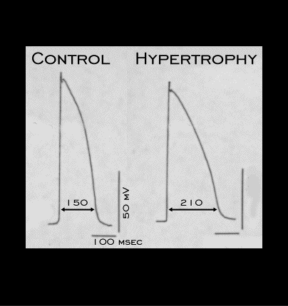
The ST segment and T wave changes associated with electrocardiographic changes induced by left ventricular hypertrophy are probably multi-factorial. They may be attributed to secondary changes, i.e. those that are secondary to the changes in magnitude, duration and sequence of the depolarizing forces. They may also be attributed to primary changes caused by abnormal voltage gradients created during repolarization. Shown here are action potentials recorded from the interventricular septum of a rabbit heart before and after left ventricular hypertrophy was induced be constriction of the aorta. Changes of this type are inhomogeneously distributed throughout the ventricle and are a likely contributor to the repolarization abnormalities.