
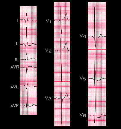
The tracing shown here is from a 28 year old male with hypertrophic obstructive cardiomyopathy (HOCM). The echocardiogram revealed a marked increase in septal and posterior wall thickness. The ECG also meets the criteria of the various left ventricular hypertrophy algorithms. Note also, the large Q waves in leads I, aVL, and V4-6. These exaggerated initial forces represent depolarization of the hypertrophied septum, i.e. are exaggerated “septal Q waves”. However, they could be mistaken for the abnormal Q waves caused by a myocardial infarction. This then is a second example of a “pseudo infarction pattern” (the prior example was in patients with ventricular pre-excitation- see page 3.5.10).