Home
Understanding the Electrocardiogram

Main navigation

  • Home
  • Understanding ECGs
    • Introduction
User account menu
  • Log in

Breadcrumb

  1. Home
  2. Understanding the Electrocardiogram In Health & Disease
  3. 3.0.0 Inter and intra-ventricular Conduction Disturbances
  4. 3.3.0 Fascicular blocks (frame 29)

3.3.17 (this is now frame 45)

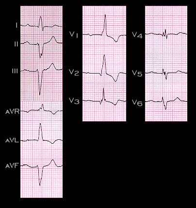
This ECG from the same patient was recorded 10 years later. It now demonstrates the development of right bundle branch block and prolongation of the PR interval to 0.28 seconds. The initial and mid portions of the QRS complex are essentially unchanged and again show the characteristics of left anterior fascicular block with the small Q in leas I and aVL and the marked left axis deviation. The Q waves in V2-V4, indicative of the prior anterior wall myocardial infarction also persist. The right bundle branch block has prolonged the duration of the QRS complex from 0.10 to 0.14 seconds and there is a prominent R’ in leads V1 and V2 and a broadened S wave in leads I, V5 and V6. Now draw the frontal plane initial, main and terminal spatial vectors and encloses them in a vector loop.

Book traversal links for 3.3.17 (this is now frame 45)

  • 3.3.16 (43)
  • Up
  • 3.3.18 (46)

Site is under construction

Book navigation

  • Introduction to First Edition
  • 1.0.0 Generation of the ECG
  • 2.0.0 The Normal Electrocardiogram
  • 3.0.0 Inter and intra-ventricular Conduction Disturbances
    • 3.0.1 Introduction Continued
    • 3.1.0 Right Bundle Branch Block (RBBB)
    • 3.2.0 Left Bundle Branch Block (LBBB)
    • 3.3.0 Fascicular blocks (frame 29)
      • 3.3.1(30)
      • 3.3.2 (31)
      • 3.3.3 (32)
      • 3.3.4 (33)
      • 3.3.5 (34)
      • 3.3.6
      • 3.3.7 (35)
      • 3.3.8 (36)
      • 3.3.9 (37)
      • 3.3.10 (38)
      • 3.3.11(39)
      • 3.3.12 (40)
      • 3.3.13 (41)
      • 3.3.14 (42)
      • 3.3.15 (new frame)
      • 3.3.16 (43)
      • 3.3.17 (this is now frame 45)
      • 3.3.18 (46)
      • 3.3.19 (47)
      • 3.3.20(48)
      • 3.3.21 (49)
      • 3.3.22(50)
      • 3.3.23 (51)
      • 3.3.24 (frame73)
      • 3.3.25 (frame 74)
    • 3.4.0 Aberrant Ventricular Conduction (52)
    • 3.5.0 Ventricular Pre-excitation (77)
  • 4.0.0 Ventricular Hypertrophy
  • 5.0.0 ELECTROLYTE ABNORMALITIES, DRUG EFFECTS AND THE LONG QT SYNDROMES
  • 6.0.0 Ischemia and Infarction - Introduction (frame i and ii)
  • 7.0.0 Tachycardias- Introduction
  • 8.0.0 The Bradycardias frame i-introduction
  • 9.0.0 The ECG of Heart Murmurs-introduction
  • 10.0.0 The Electrocardiogram in the Emergency Department-Introduction
Powered by Drupal