Home
Understanding the Electrocardiogram

Main navigation

  • Home
  • Understanding ECGs
    • Introduction
User account menu
  • Log in

Breadcrumb

  1. Home
  2. Understanding the Electrocardiogram In Health & Disease
  3. 3.0.0 Inter and intra-ventricular Conduction Disturbances
  4. 3.3.0 Fascicular blocks (frame 29)

3.3.8 (36)

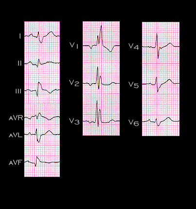
The ECG shown here demonstrates right bundle branch block with left posterior fascicular block. The left posterior fascicular block is manifested by the initial small R wave in leads I and aVL and small Q in leads II, and aVF. These are followed by the larger S waves in leads I and aVL and the R waves in II, III and aVF. The right bundle branch block is manifested by the increased duration of these terminal forces and by the broad R’ in leads aVR, V1, V2 and V3. The duration of the QRS complex is greater than 0.120 second, measuring approximately 0.160 seconds (160 ms). Try to draw the initial, main and terminal QRS spatial vectors in the frontal plane. Then draw the vector loop that encompasses these vectors.

Book traversal links for 3.3.8 (36)

  • 3.3.7 (35)
  • Up
  • 3.3.9 (37)

Site is under construction

Book navigation

  • Introduction to First Edition
  • 1.0.0 Generation of the ECG
  • 2.0.0 The Normal Electrocardiogram
  • 3.0.0 Inter and intra-ventricular Conduction Disturbances
    • 3.0.1 Introduction Continued
    • 3.1.0 Right Bundle Branch Block (RBBB)
    • 3.2.0 Left Bundle Branch Block (LBBB)
    • 3.3.0 Fascicular blocks (frame 29)
      • 3.3.1(30)
      • 3.3.2 (31)
      • 3.3.3 (32)
      • 3.3.4 (33)
      • 3.3.5 (34)
      • 3.3.6
      • 3.3.7 (35)
      • 3.3.8 (36)
      • 3.3.9 (37)
      • 3.3.10 (38)
      • 3.3.11(39)
      • 3.3.12 (40)
      • 3.3.13 (41)
      • 3.3.14 (42)
      • 3.3.15 (new frame)
      • 3.3.16 (43)
      • 3.3.17 (this is now frame 45)
      • 3.3.18 (46)
      • 3.3.19 (47)
      • 3.3.20(48)
      • 3.3.21 (49)
      • 3.3.22(50)
      • 3.3.23 (51)
      • 3.3.24 (frame73)
      • 3.3.25 (frame 74)
    • 3.4.0 Aberrant Ventricular Conduction (52)
    • 3.5.0 Ventricular Pre-excitation (77)
  • 4.0.0 Ventricular Hypertrophy
  • 5.0.0 ELECTROLYTE ABNORMALITIES, DRUG EFFECTS AND THE LONG QT SYNDROMES
  • 6.0.0 Ischemia and Infarction - Introduction (frame i and ii)
  • 7.0.0 Tachycardias- Introduction
  • 8.0.0 The Bradycardias frame i-introduction
  • 9.0.0 The ECG of Heart Murmurs-introduction
  • 10.0.0 The Electrocardiogram in the Emergency Department-Introduction
Powered by Drupal