
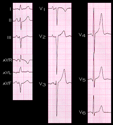
This ECG from a 61 year old patient demonstrates the pertinent features of left anterior fascicular block. A small Q wave is present in leads I, aVL, V5 and V6 and there is a small R wave in leads 3 and aVF. The frontal plane axis is -70 degrees and the duration of the QRS complex is 0.10 seconds. Plotting the initial and main QRS spatial vectors in the frontal plane, as demonstrated on the next page, is a useful way to visualize the features of this conduction disturbance.