Home
Understanding the Electrocardiogram

Main navigation

  • Home
  • Understanding ECGs
    • Introduction
User account menu
  • Log in

Breadcrumb

  1. Home
  2. Understanding the Electrocardiogram In Health & Disease
  3. 3.0.0 Inter and intra-ventricular Conduction Disturbances
  4. 3.3.0 Fascicular blocks (frame 29)

3.3.10 (38)

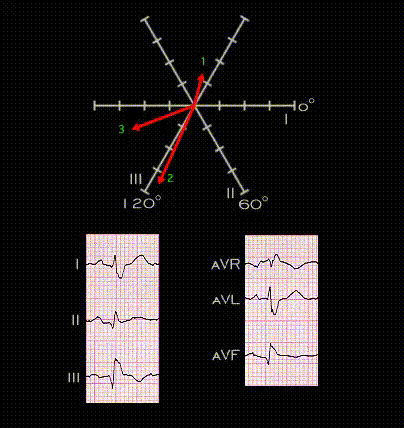
The vector loop shown here rotates in a clockwise direction to encompass these three spatial vectors. It demonstrates the superior direction of the initial vector, the right axis deviation of the main QRS vector and the slowed terminal component caused by the right bundle branch block. The combination of right bundle branch block and right fascicular block is an example of bifascicular block, so named because if represents slowing of conduction or block in more than one fascicle of the conduction system

Book traversal links for 3.3.10 (38)

  • 3.3.9 (37)
  • Up
  • 3.3.11(39)

Site is under construction

Book navigation

  • Introduction to First Edition
  • 1.0.0 Generation of the ECG
  • 2.0.0 The Normal Electrocardiogram
  • 3.0.0 Inter and intra-ventricular Conduction Disturbances
    • 3.0.1 Introduction Continued
    • 3.1.0 Right Bundle Branch Block (RBBB)
    • 3.2.0 Left Bundle Branch Block (LBBB)
    • 3.3.0 Fascicular blocks (frame 29)
      • 3.3.1(30)
      • 3.3.2 (31)
      • 3.3.3 (32)
      • 3.3.4 (33)
      • 3.3.5 (34)
      • 3.3.6
      • 3.3.7 (35)
      • 3.3.8 (36)
      • 3.3.9 (37)
      • 3.3.10 (38)
      • 3.3.11(39)
      • 3.3.12 (40)
      • 3.3.13 (41)
      • 3.3.14 (42)
      • 3.3.15 (new frame)
      • 3.3.16 (43)
      • 3.3.17 (this is now frame 45)
      • 3.3.18 (46)
      • 3.3.19 (47)
      • 3.3.20(48)
      • 3.3.21 (49)
      • 3.3.22(50)
      • 3.3.23 (51)
      • 3.3.24 (frame73)
      • 3.3.25 (frame 74)
    • 3.4.0 Aberrant Ventricular Conduction (52)
    • 3.5.0 Ventricular Pre-excitation (77)
  • 4.0.0 Ventricular Hypertrophy
  • 5.0.0 ELECTROLYTE ABNORMALITIES, DRUG EFFECTS AND THE LONG QT SYNDROMES
  • 6.0.0 Ischemia and Infarction - Introduction (frame i and ii)
  • 7.0.0 Tachycardias- Introduction
  • 8.0.0 The Bradycardias frame i-introduction
  • 9.0.0 The ECG of Heart Murmurs-introduction
  • 10.0.0 The Electrocardiogram in the Emergency Department-Introduction
Powered by Drupal