Home
Understanding the Electrocardiogram

Main navigation

  • Home
  • Understanding ECGs
    • Introduction
User account menu
  • Log in

Breadcrumb

  1. Home
  2. Understanding the Electrocardiogram In Health & Disease
  3. 8.0.0 The Bradycardias frame i-introduction
  4. 8.1.0 Sinus Bradycardia and AV block (frame 1)
  5. 8.1.20 (frame 21)

8.1.29 (30)

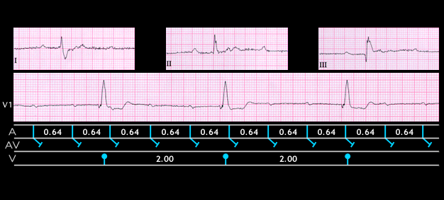
The more distal the site of the escape pacemaker, the slower its rate and the more abnormal the QRS complex. This tracing was recorded from a 54 year old male following an episode of syncope. The atrial rate is 90 (P-P intrval = 0.64sec), the ventricular rate is 30 (R-R interval  = 2.00 sec) and there is complete AV block with AV dissociation. The QRS complex is 0.16 seconds in duration and shows right bundle branch block. This indicates that the escape pacemaker is located in the left ventricle. Syncope occurring in the setting of high grade or complete AV block is referred to as Stokes-Adams disease. The syncope occurs as the result of ventricular tachycardia or even more profound bradycardia. This scenrio is prevented by ventricular pacing

Book traversal links for 8.1.29 (30)

  • 8.1.28 (29)
  • Up
  • 8.1.30 (frame 31)

Site is under construction

Book navigation

  • Introduction to First Edition
  • 1.0.0 Generation of the ECG
  • 2.0.0 The Normal Electrocardiogram
  • 3.0.0 Inter and intra-ventricular Conduction Disturbances
  • 4.0.0 Ventricular Hypertrophy
  • 5.0.0 ELECTROLYTE ABNORMALITIES, DRUG EFFECTS AND THE LONG QT SYNDROMES
  • 6.0.0 Ischemia and Infarction - Introduction (frame i and ii)
  • 7.0.0 Tachycardias- Introduction
  • 8.0.0 The Bradycardias frame i-introduction
    • 8.1.0 Sinus Bradycardia and AV block (frame 1)
      • 8.1.1 (2)
      • 8.1.2 (3)
      • 8.1.3 (4)
      • 8.1.4 (5)
      • 8.1.5 (6)
      • 8.1.6 (7)
      • 8.1.7 (8)
      • 8.1.8 (9)
      • 8.1.9 (10)
      • 8.1.10 (frame 11)
      • 8.1.20 (frame 21)
        • 8.1.21 (22)
        • 8.1.22 (23)
        • 8.1.23 (24)
        • 8.1.24 (25)
        • 8.1.25 (26)
        • 8.1.26 (27)
        • 8.1.27 (28)
        • 8.1.28 (29)
        • 8.1.29 (30)
      • 8.1.30 (frame 31)
    • 8.2.0 SA Block (frame 41)
    • 8.3.0 Group Beating (frame 57)
  • 9.0.0 The ECG of Heart Murmurs-introduction
  • 10.0.0 The Electrocardiogram in the Emergency Department-Introduction
Powered by Drupal