Home
Understanding the Electrocardiogram

Main navigation

  • Home
  • Understanding ECGs
    • Introduction
User account menu
  • Log in

Breadcrumb

  1. Home
  2. Understanding the Electrocardiogram In Health & Disease
  3. 8.0.0 The Bradycardias frame i-introduction
  4. 8.1.0 Sinus Bradycardia and AV block (frame 1)
  5. 8.1.20 (frame 21)

8.1.23 (24)

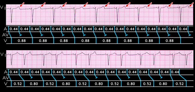
When the atrial rate is rapid and there is no evidence of distal conduction system disease, such as fascicular or bundle branch block, the site of 2:1 AV block is more likely to be within the AV node than distal to it.

In both tracings displayed here, the atrial rate is 140. In the upper tracing, there is 2:1 AV conduction but the blocked P waves (arrows) are difficult to see because they fall within the T wave. The lower strip was recorded the next day and shows type I 2nd degree AV block with 3:2 conduction. This suggests that the site of block associated with the 2:1 conduction was also within the AV node.

Book traversal links for 8.1.23 (24)

  • 8.1.22 (23)
  • Up
  • 8.1.24 (25)

Site is under construction

Book navigation

  • Introduction to First Edition
  • 1.0.0 Generation of the ECG
  • 2.0.0 The Normal Electrocardiogram
  • 3.0.0 Inter and intra-ventricular Conduction Disturbances
  • 4.0.0 Ventricular Hypertrophy
  • 5.0.0 ELECTROLYTE ABNORMALITIES, DRUG EFFECTS AND THE LONG QT SYNDROMES
  • 6.0.0 Ischemia and Infarction - Introduction (frame i and ii)
  • 7.0.0 Tachycardias- Introduction
  • 8.0.0 The Bradycardias frame i-introduction
    • 8.1.0 Sinus Bradycardia and AV block (frame 1)
      • 8.1.1 (2)
      • 8.1.2 (3)
      • 8.1.3 (4)
      • 8.1.4 (5)
      • 8.1.5 (6)
      • 8.1.6 (7)
      • 8.1.7 (8)
      • 8.1.8 (9)
      • 8.1.9 (10)
      • 8.1.10 (frame 11)
      • 8.1.20 (frame 21)
        • 8.1.21 (22)
        • 8.1.22 (23)
        • 8.1.23 (24)
        • 8.1.24 (25)
        • 8.1.25 (26)
        • 8.1.26 (27)
        • 8.1.27 (28)
        • 8.1.28 (29)
        • 8.1.29 (30)
      • 8.1.30 (frame 31)
    • 8.2.0 SA Block (frame 41)
    • 8.3.0 Group Beating (frame 57)
  • 9.0.0 The ECG of Heart Murmurs-introduction
  • 10.0.0 The Electrocardiogram in the Emergency Department-Introduction
Powered by Drupal