Home
Understanding the Electrocardiogram

Main navigation

  • Home
  • Understanding ECGs
    • Introduction
User account menu
  • Log in

Breadcrumb

  1. Home
  2. Understanding the Electrocardiogram In Health & Disease
  3. 8.0.0 The Bradycardias frame i-introduction

8.2.0 SA Block (frame 41)

The electrocardiographic determination of AV conduction disturbances is reasonably straight forward because the markers of atrial and ventricular depolarization (the P wave and QRS complex ) can be detected on the body surface and recorded on the standard body surface electrocardiogram. In contrast, the ECG determination of disturbances in conduction from the sinus node to the surrounding atrial myocardium, i.e. sino-atrial (SA) conduction disturbances, is more difficult because the electrical signal caused by sinus node depolarization is not strong enough to be recorded on the body surface. For this reason, sinus node activity must be deduced from the rate and rhythm of P waves that originate in the sinus node.

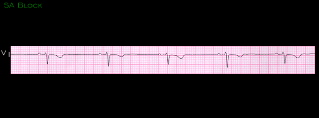
This ECG is from a 23 year old female with no known heart disease. Try to determine where sinus node depolarization occurs relative to the P wave.

  • 8.2.1 (42)
  • 8.2.2 (43)
  • 8.2.3(44)
  • 8.2.4 (45)
  • 8.2.5 (46)
  • 8.2.6 (47)
  • 8.2.7 (48)
  • 8.2.8 (49)
  • 8.2.9 (50)
  • 8.2.10 (frame 51)
  • 8.2.11 (52)
  • 8.2.12 (53)
  • 8.2.13 (54)
  • 8.2.14 (55)
  • 8.2.15 (56)

Book traversal links for 8.2.0 SA Block (frame 41)

  • 8.1.39 (40)
  • Up
  • 8.2.1 (42)

Site is under construction

Book navigation

  • Introduction to First Edition
  • 1.0.0 Generation of the ECG
  • 2.0.0 The Normal Electrocardiogram
  • 3.0.0 Inter and intra-ventricular Conduction Disturbances
  • 4.0.0 Ventricular Hypertrophy
  • 5.0.0 ELECTROLYTE ABNORMALITIES, DRUG EFFECTS AND THE LONG QT SYNDROMES
  • 6.0.0 Ischemia and Infarction - Introduction (frame i and ii)
  • 7.0.0 Tachycardias- Introduction
  • 8.0.0 The Bradycardias frame i-introduction
    • 8.1.0 Sinus Bradycardia and AV block (frame 1)
    • 8.2.0 SA Block (frame 41)
      • 8.2.1 (42)
      • 8.2.2 (43)
      • 8.2.3(44)
      • 8.2.4 (45)
      • 8.2.5 (46)
      • 8.2.6 (47)
      • 8.2.7 (48)
      • 8.2.8 (49)
      • 8.2.9 (50)
      • 8.2.10 (frame 51)
      • 8.2.11 (52)
      • 8.2.12 (53)
      • 8.2.13 (54)
      • 8.2.14 (55)
      • 8.2.15 (56)
    • 8.3.0 Group Beating (frame 57)
  • 9.0.0 The ECG of Heart Murmurs-introduction
  • 10.0.0 The Electrocardiogram in the Emergency Department-Introduction
Powered by Drupal