Home
Understanding the Electrocardiogram

Main navigation

  • Home
  • Understanding ECGs
    • Introduction
User account menu
  • Log in

Breadcrumb

  1. Home
  2. Understanding the Electrocardiogram In Health & Disease
  3. 8.0.0 The Bradycardias frame i-introduction
  4. 8.1.0 Sinus Bradycardia and AV block (frame 1)

8.1.30 (frame 31)

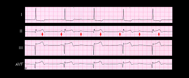
Conduction delay within the AV node, causing first or second degree and/or high grade AV block frequently accompanies an acute inferior wall myocardial infarction and is almost always transient. The frontal plane (limb) leads shown here are from an 83 year old female with an acute inferior wall infarction. There is complete AV block with AV dissociation and a narrow QRS complex escape rhythm, indicating that the site of block is within the AV node and that the escape pacemaker is located above the His bifurcation. Its rate is 44 beats per minute (R-R interval = 1.36 sec). The P waves are best seen in lead II (arrows). They have a normal configuration and a rate of 67 (P-P interval = 0.9 sec). The Q waves in leads II, III and aVF and the ST segment elevation in these leads reflect the acute inferior wall infarction.

  • 8.1.31 (32)
  • 8.1.32 (33)
  • 8.1.33 (34)
  • 8.1.34 (35)
  • 8.1.35 (37-this is correct)
  • 8.1.36 (36)
  • 8.1.37 (38)
  • 8.1.38 (39)
  • 8.1.39 (40)

Book traversal links for 8.1.30 (frame 31)

  • 8.1.29 (30)
  • Up
  • 8.1.31 (32)

Site is under construction

Book navigation

  • Introduction to First Edition
  • 1.0.0 Generation of the ECG
  • 2.0.0 The Normal Electrocardiogram
  • 3.0.0 Inter and intra-ventricular Conduction Disturbances
  • 4.0.0 Ventricular Hypertrophy
  • 5.0.0 ELECTROLYTE ABNORMALITIES, DRUG EFFECTS AND THE LONG QT SYNDROMES
  • 6.0.0 Ischemia and Infarction - Introduction (frame i and ii)
  • 7.0.0 Tachycardias- Introduction
  • 8.0.0 The Bradycardias frame i-introduction
    • 8.1.0 Sinus Bradycardia and AV block (frame 1)
      • 8.1.1 (2)
      • 8.1.2 (3)
      • 8.1.3 (4)
      • 8.1.4 (5)
      • 8.1.5 (6)
      • 8.1.6 (7)
      • 8.1.7 (8)
      • 8.1.8 (9)
      • 8.1.9 (10)
      • 8.1.10 (frame 11)
      • 8.1.20 (frame 21)
      • 8.1.30 (frame 31)
        • 8.1.31 (32)
        • 8.1.32 (33)
        • 8.1.33 (34)
        • 8.1.34 (35)
        • 8.1.35 (37-this is correct)
        • 8.1.36 (36)
        • 8.1.37 (38)
        • 8.1.38 (39)
        • 8.1.39 (40)
    • 8.2.0 SA Block (frame 41)
    • 8.3.0 Group Beating (frame 57)
  • 9.0.0 The ECG of Heart Murmurs-introduction
  • 10.0.0 The Electrocardiogram in the Emergency Department-Introduction
Powered by Drupal