
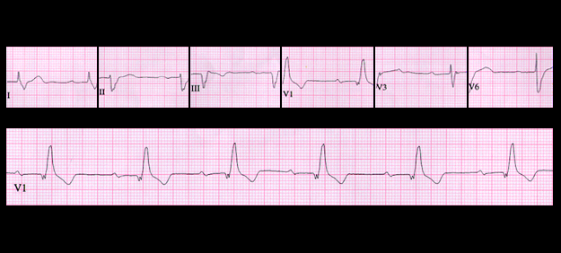
This tracing is from a 74 year old male with hypertension and heart failure. The rate is 48, the P waves appear normal, there is 1:1 AV conduction and the PR interval is prolonged to approximately 0.32 seconds. Note that there is also right bundle branch block with left axis deviation (-60 degrees) of the initial portion of the QRS complex. This indicates that there is block in the anterior fascicle of the left bundle as well as block in the right bundle. The PR prolongation may be due to slow conduction in the AV node and/or in the left posterior fascicle. This ECG suggests diffuse conducting system disease and is an harbinger of higher degrees of AV block, including complete AV block.