Home
Understanding the Electrocardiogram

Main navigation

  • Home
  • Understanding ECGs
    • Introduction
User account menu
  • Log in

Breadcrumb

  1. Home
  2. Understanding the Electrocardiogram In Health & Disease
  3. 8.0.0 The Bradycardias frame i-introduction
  4. 8.1.0 Sinus Bradycardia and AV block (frame 1)

8.1.10 (frame 11)

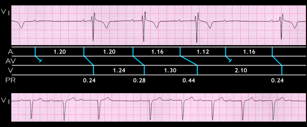
[SHOW JUST THE TOP STRIP AND LADDER DIAGRAM] The ECG strip and accompanying ladder diagram shown here is an example of Mobitz type I 2nd degree AV block with Wenckebach periodicity. It is from a 25 year old male who feinted after donating a pint of blood. There is sinus bradycardias (rate of 50), progressive prolongation of the PR interval (0.24, 0.28 and 0.44 seconds) folowed by a P wave that is blocked within th AV junction. This results in 4:3 AV block. Note that the RR interval encompassing the blocked P wave of 2.10 seconds. is less than twice the preceding RR interval (2 x 1.3 =2.6 seconds) This is characteristic of type I 2nd degree AV block which, in this patient, was due to an increase in vagal tone associated with the phlebotomy.

  • 8.1.11 (12)
  • 8.1.12 (13)
  • 8.1.13 (14)
  • 8.1.14 (15)
  • 8.1.15 (16)
  • 8.1.16 (17)
  • 8.1.17 (18)
  • 8.1.18 (19)
  • 8.1.19 (frame 20)

Book traversal links for 8.1.10 (frame 11)

  • 8.1.9 (10)
  • Up
  • 8.1.11 (12)

Site is under construction

Book navigation

  • Introduction to First Edition
  • 1.0.0 Generation of the ECG
  • 2.0.0 The Normal Electrocardiogram
  • 3.0.0 Inter and intra-ventricular Conduction Disturbances
  • 4.0.0 Ventricular Hypertrophy
  • 5.0.0 ELECTROLYTE ABNORMALITIES, DRUG EFFECTS AND THE LONG QT SYNDROMES
  • 6.0.0 Ischemia and Infarction - Introduction (frame i and ii)
  • 7.0.0 Tachycardias- Introduction
  • 8.0.0 The Bradycardias frame i-introduction
    • 8.1.0 Sinus Bradycardia and AV block (frame 1)
      • 8.1.1 (2)
      • 8.1.2 (3)
      • 8.1.3 (4)
      • 8.1.4 (5)
      • 8.1.5 (6)
      • 8.1.6 (7)
      • 8.1.7 (8)
      • 8.1.8 (9)
      • 8.1.9 (10)
      • 8.1.10 (frame 11)
        • 8.1.11 (12)
        • 8.1.12 (13)
        • 8.1.13 (14)
        • 8.1.14 (15)
        • 8.1.15 (16)
        • 8.1.16 (17)
        • 8.1.17 (18)
        • 8.1.18 (19)
        • 8.1.19 (frame 20)
      • 8.1.20 (frame 21)
      • 8.1.30 (frame 31)
    • 8.2.0 SA Block (frame 41)
    • 8.3.0 Group Beating (frame 57)
  • 9.0.0 The ECG of Heart Murmurs-introduction
  • 10.0.0 The Electrocardiogram in the Emergency Department-Introduction
Powered by Drupal