
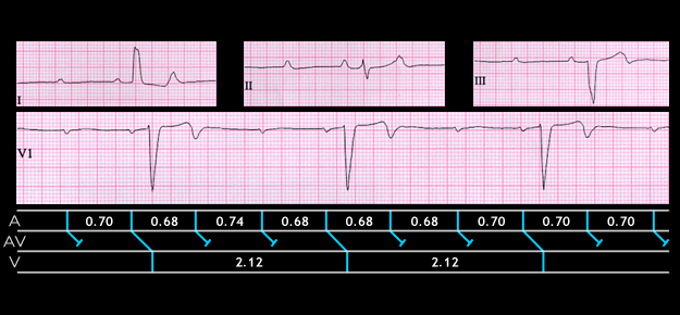
Rarely, only very third P wave is conducted. This is referred to as high grade AV block and is illustrated in the ECG shown here. The tracing is from a 47 year old female. The atrial rate is 85 and there is 3:1 AV block. The QRS complexes reveal left bundle branch block. This finding, in combination with the AV block, suggests diffuse conducting system disease.