Home
Understanding the Electrocardiogram

Main navigation

  • Home
  • Understanding ECGs
    • Introduction
User account menu
  • Log in

Breadcrumb

  1. Home
  2. Understanding the Electrocardiogram In Health & Disease
  3. 8.0.0 The Bradycardias frame i-introduction
  4. 8.1.0 Sinus Bradycardia and AV block (frame 1)

8.1.3 (4)

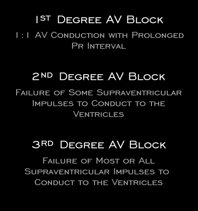
Normally, there is a 1:1 relationship between the P waves and the QRS complexes. The PR interval, which includes inter and intra-atrial, AV nodal and His-Purkinje conduction, has a normal duration of 0.12 to 2.0 seconds. Conduction slowing within the AV node and His-Purkinje system, which together comprise the AV junction, is manifest by prolongation of the PR interval beyond 0.20 seconds and/or the failure of some or all of the atrial impulses to conduct to the ventricles. Prolongation of the PR interval beyond 0.20 seconds is referred to as 1st degree AV block, although some readers refer to it merely as “prolonged PR interval.” Failure of some, but not all of the atrial impulses to propagate to the ventricles is referred to as 2nd degree AV block, and failure of most or all of the impulses to propagate to the ventricles is variously referred to as high grade, 3rd degree, or complete AV block.

Book traversal links for 8.1.3 (4)

  • 8.1.2 (3)
  • Up
  • 8.1.4 (5)

Site is under construction

Book navigation

  • Introduction to First Edition
  • 1.0.0 Generation of the ECG
  • 2.0.0 The Normal Electrocardiogram
  • 3.0.0 Inter and intra-ventricular Conduction Disturbances
  • 4.0.0 Ventricular Hypertrophy
  • 5.0.0 ELECTROLYTE ABNORMALITIES, DRUG EFFECTS AND THE LONG QT SYNDROMES
  • 6.0.0 Ischemia and Infarction - Introduction (frame i and ii)
  • 7.0.0 Tachycardias- Introduction
  • 8.0.0 The Bradycardias frame i-introduction
    • 8.1.0 Sinus Bradycardia and AV block (frame 1)
      • 8.1.1 (2)
      • 8.1.2 (3)
      • 8.1.3 (4)
      • 8.1.4 (5)
      • 8.1.5 (6)
      • 8.1.6 (7)
      • 8.1.7 (8)
      • 8.1.8 (9)
      • 8.1.9 (10)
      • 8.1.10 (frame 11)
      • 8.1.20 (frame 21)
      • 8.1.30 (frame 31)
    • 8.2.0 SA Block (frame 41)
    • 8.3.0 Group Beating (frame 57)
  • 9.0.0 The ECG of Heart Murmurs-introduction
  • 10.0.0 The Electrocardiogram in the Emergency Department-Introduction
Powered by Drupal