Home
Understanding the Electrocardiogram

Main navigation

  • Home
  • Understanding ECGs
    • Introduction
User account menu
  • Log in

Breadcrumb

  1. Home
  2. Understanding the Electrocardiogram In Health & Disease
  3. 8.0.0 The Bradycardias frame i-introduction
  4. 8.1.0 Sinus Bradycardia and AV block (frame 1)
  5. 8.1.20 (frame 21)

8.1.21 (22)

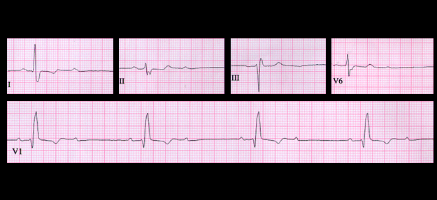
When there is 2:1 AV block, it is often impossible to determine from the ECG whether the site of block is within the AV node or the more distal conduction system since the characteristics of the PR intervals prior to the blocked P wave can not be established. This ECG shows sinus rhythm with an atrial rate of 68 (PP interval = 0.88 seconds) and 2:1 AV block. There is right bundle branch block with left axis deviation, and the PR interval of the conducted P waves is 0.20 seconds. Although the tracing suggests diffuse conducting system disease, it is impossible to determine the site of block of the non-conducted P waves from this ECG.

The tracing from the next day, shown the following page, helps to identify the site of block.

Book traversal links for 8.1.21 (22)

  • 8.1.20 (frame 21)
  • Up
  • 8.1.22 (23)

Site is under construction

Book navigation

  • Introduction to First Edition
  • 1.0.0 Generation of the ECG
  • 2.0.0 The Normal Electrocardiogram
  • 3.0.0 Inter and intra-ventricular Conduction Disturbances
  • 4.0.0 Ventricular Hypertrophy
  • 5.0.0 ELECTROLYTE ABNORMALITIES, DRUG EFFECTS AND THE LONG QT SYNDROMES
  • 6.0.0 Ischemia and Infarction - Introduction (frame i and ii)
  • 7.0.0 Tachycardias- Introduction
  • 8.0.0 The Bradycardias frame i-introduction
    • 8.1.0 Sinus Bradycardia and AV block (frame 1)
      • 8.1.1 (2)
      • 8.1.2 (3)
      • 8.1.3 (4)
      • 8.1.4 (5)
      • 8.1.5 (6)
      • 8.1.6 (7)
      • 8.1.7 (8)
      • 8.1.8 (9)
      • 8.1.9 (10)
      • 8.1.10 (frame 11)
      • 8.1.20 (frame 21)
        • 8.1.21 (22)
        • 8.1.22 (23)
        • 8.1.23 (24)
        • 8.1.24 (25)
        • 8.1.25 (26)
        • 8.1.26 (27)
        • 8.1.27 (28)
        • 8.1.28 (29)
        • 8.1.29 (30)
      • 8.1.30 (frame 31)
    • 8.2.0 SA Block (frame 41)
    • 8.3.0 Group Beating (frame 57)
  • 9.0.0 The ECG of Heart Murmurs-introduction
  • 10.0.0 The Electrocardiogram in the Emergency Department-Introduction
Powered by Drupal