
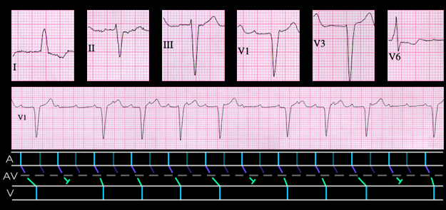
Two levels of block within the AV node may also occur when the atrial rate is less than that usually encountered in atrial flutter. The ECG shown here is from a 54 year old female with diffuse connective tissue disease and left bundle branch block. The rhythm is ectopic atrial tachycardia with an atrial rate of 175. In the segment shown here, there is group beating with a group of four QRS complexes followed by a group of three and a P:QRS relationship that is either 2:1 or 4:1. As illustrated in the ladder diagram, the rhythm is explained by postulating 2 levels of AV nodal block with fixed 2:1 block in the upper portion of the node and then 5:4 followed by 4:3 block in the lower.