Home
Understanding the Electrocardiogram

Main navigation

  • Home
  • Understanding ECGs
    • Introduction
User account menu
  • Log in

Breadcrumb

  1. Home
  2. Understanding the Electrocardiogram In Health & Disease
  3. 8.0.0 The Bradycardias frame i-introduction
  4. 8.1.0 Sinus Bradycardia and AV block (frame 1)
  5. 8.1.10 (frame 11)

8.1.13 (14)

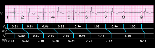
The ECG displayed here is from a 24 year old highly trained marathon runner with no evidence of heart disease. It illustrates an atypical form of type 1 2nd degree AV block in which there is continuous variability in the PP, PR and RR intervals.

The PP interval between beats 1 and 2 and between beats 2 and 3 measures 0.84 seconds. It then lengthens to 1.08 seconds between beats 6 and 7.

The PR interval shortens from 0.38 seconds in beat 1 to 0.22 seconds in beat 6. It then lengthens to 0.32 seconds in beat 7 and the 8th P wave is blocked. The next P wave (#9) conducts with a PR interval of 0.16 seconds.

These changes in sinus rate and AV conduction reflect the phasic variations in autonomic balance present in younger individuals and in highly trained athletes.

Book traversal links for 8.1.13 (14)

  • 8.1.12 (13)
  • Up
  • 8.1.14 (15)

Site is under construction

Book navigation

  • Introduction to First Edition
  • 1.0.0 Generation of the ECG
  • 2.0.0 The Normal Electrocardiogram
  • 3.0.0 Inter and intra-ventricular Conduction Disturbances
  • 4.0.0 Ventricular Hypertrophy
  • 5.0.0 ELECTROLYTE ABNORMALITIES, DRUG EFFECTS AND THE LONG QT SYNDROMES
  • 6.0.0 Ischemia and Infarction - Introduction (frame i and ii)
  • 7.0.0 Tachycardias- Introduction
  • 8.0.0 The Bradycardias frame i-introduction
    • 8.1.0 Sinus Bradycardia and AV block (frame 1)
      • 8.1.1 (2)
      • 8.1.2 (3)
      • 8.1.3 (4)
      • 8.1.4 (5)
      • 8.1.5 (6)
      • 8.1.6 (7)
      • 8.1.7 (8)
      • 8.1.8 (9)
      • 8.1.9 (10)
      • 8.1.10 (frame 11)
        • 8.1.11 (12)
        • 8.1.12 (13)
        • 8.1.13 (14)
        • 8.1.14 (15)
        • 8.1.15 (16)
        • 8.1.16 (17)
        • 8.1.17 (18)
        • 8.1.18 (19)
        • 8.1.19 (frame 20)
      • 8.1.20 (frame 21)
      • 8.1.30 (frame 31)
    • 8.2.0 SA Block (frame 41)
    • 8.3.0 Group Beating (frame 57)
  • 9.0.0 The ECG of Heart Murmurs-introduction
  • 10.0.0 The Electrocardiogram in the Emergency Department-Introduction
Powered by Drupal