Home
Understanding the Electrocardiogram

Main navigation

  • Home
  • Understanding ECGs
    • Introduction
User account menu
  • Log in

Breadcrumb

  1. Home
  2. Understanding the Electrocardiogram In Health & Disease
  3. 1.0.0 Generation of the ECG
  4. 1.8.0 Electrodes

1.8.9

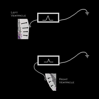
Extending this concept one step further, consider the heart with its dissimilar ventricular dimensions. If the ventricles were to be isolated and a signal recorded by a unipolar electrode located on the epicardial surface of each ventricle as it is depolarized from the endocardium to the epicardium, a positive wave form would be generated from each location. However, that recorded over the left ventricle would be of greater magnitude than that generated from the right ventricle because of the greater muscle mass of the left ventricle.

Book traversal links for 1.8.9

  • 1.8.8
  • Up
  • 1.8.10

Site is under construction

Book navigation

  • Introduction to First Edition
  • 1.0.0 Generation of the ECG
    • 1.1.0 Basic Physiology
    • 1.2.0 SA Node & Pacemakers
    • 1.3.0 Atrial Depolarization
    • 1.4.0 Atrio Ventricular Conduction
    • 1.5.0 Ventricular Depolarization
    • 1.6.0 Repolarization
    • 1.7.0 Events at the Cellular Level
    • 1.8.0 Electrodes
      • 1.8.1
      • 1.8.2
      • 1.8.3
      • 1.8.4
      • 1.8.5
      • 1.8.6
      • 1.8.7
      • 1.8.8
      • 1.8.9
      • 1.8.10
      • 1.8.11
      • 1.8.12
      • 1.8.13
      • 1.8.14
    • 1.9.0 ECG Leads and Placement
  • 2.0.0 The Normal Electrocardiogram
  • 3.0.0 Inter and intra-ventricular Conduction Disturbances
  • 4.0.0 Ventricular Hypertrophy
  • 5.0.0 ELECTROLYTE ABNORMALITIES, DRUG EFFECTS AND THE LONG QT SYNDROMES
  • 6.0.0 Ischemia and Infarction - Introduction (frame i and ii)
  • 7.0.0 Tachycardias- Introduction
  • 8.0.0 The Bradycardias frame i-introduction
  • 9.0.0 The ECG of Heart Murmurs-introduction
  • 10.0.0 The Electrocardiogram in the Emergency Department-Introduction
Powered by Drupal