Home
Understanding the Electrocardiogram

Main navigation

  • Home
  • Understanding ECGs
    • Introduction
User account menu
  • Log in

Breadcrumb

  1. Home
  2. Understanding the Electrocardiogram In Health & Disease
  3. 8.0.0 The Bradycardias frame i-introduction
  4. 8.1.0 Sinus Bradycardia and AV block (frame 1)
  5. 8.1.30 (frame 31)

8.1.39 (40)

When there is AV dissociation and the atrial rate is more rapid than the escape rhythm, the AV dissociation is invariably the result of AV block. When the rate of an AV junctional or ventricular rhythm is more rapid than the atrial rate, there will also be AV dissociation but in this setting, P waves will not conduct to the ventricles because the AV node will be refractory. If P waves do occur when the AV is no longer refractory, they will conduct and interfere with the AV dissociation, This rhythm is referred to as AV dissociation with interference and was discussed in the previous chapter (see pages 7.2.19 and 7.2.20).

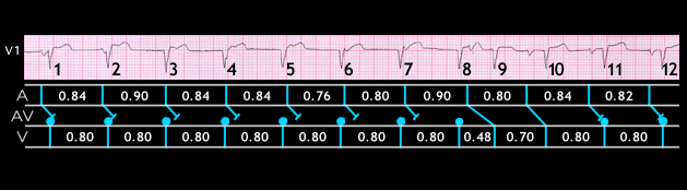
The ECG shown here is another example of this rhythm. The QRS complexes numbered 1-8 are the result of an accelerated junctional rhythm with a rate of 75 BPM (R - R interval = 0.8 seconds). The atrial rate is slightly slower, and the first 7 P waves do not conduct because the AV node is refractory, The 8th P wave occurs just after the 8th QRS complex. The AV junction has now partially recovered its ability to conduct and this P wave conducts to the ventricle with a prolonged PR interval (0.32 seconds) causing QRS complex #9. As a result the RR interval between QRS complexes #8 and 9 is only 0.48 seconds. The next P wave is also able to conduct to the ventricles, this time with a PR interval of 0.24 seconds, causing QRS complex #10. The last 2 QRS complexes (#s 11 and 12) are again the result of the accelerated AV junctional rhythm.

Book traversal links for 8.1.39 (40)

  • 8.1.38 (39)
  • Up
  • 8.2.0 SA Block (frame 41)

Site is under construction

Book navigation

  • Introduction to First Edition
  • 1.0.0 Generation of the ECG
  • 2.0.0 The Normal Electrocardiogram
  • 3.0.0 Inter and intra-ventricular Conduction Disturbances
  • 4.0.0 Ventricular Hypertrophy
  • 5.0.0 ELECTROLYTE ABNORMALITIES, DRUG EFFECTS AND THE LONG QT SYNDROMES
  • 6.0.0 Ischemia and Infarction - Introduction (frame i and ii)
  • 7.0.0 Tachycardias- Introduction
  • 8.0.0 The Bradycardias frame i-introduction
    • 8.1.0 Sinus Bradycardia and AV block (frame 1)
      • 8.1.1 (2)
      • 8.1.2 (3)
      • 8.1.3 (4)
      • 8.1.4 (5)
      • 8.1.5 (6)
      • 8.1.6 (7)
      • 8.1.7 (8)
      • 8.1.8 (9)
      • 8.1.9 (10)
      • 8.1.10 (frame 11)
      • 8.1.20 (frame 21)
      • 8.1.30 (frame 31)
        • 8.1.31 (32)
        • 8.1.32 (33)
        • 8.1.33 (34)
        • 8.1.34 (35)
        • 8.1.35 (37-this is correct)
        • 8.1.36 (36)
        • 8.1.37 (38)
        • 8.1.38 (39)
        • 8.1.39 (40)
    • 8.2.0 SA Block (frame 41)
    • 8.3.0 Group Beating (frame 57)
  • 9.0.0 The ECG of Heart Murmurs-introduction
  • 10.0.0 The Electrocardiogram in the Emergency Department-Introduction
Powered by Drupal