Home
Understanding the Electrocardiogram

Main navigation

  • Home
  • Understanding ECGs
    • Introduction
User account menu
  • Log in

Breadcrumb

  1. Home
  2. Understanding the Electrocardiogram In Health & Disease
  3. 6.0.0 Ischemia and Infarction - Introduction (frame i and ii)
  4. 6.5.0 (frame 106) FALSE POSITIVES
  5. 6.5.30 (frame 136)

6.5.31 (137)

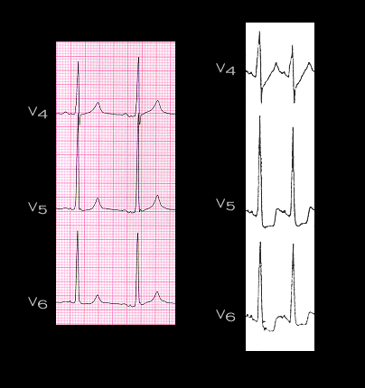
In patients with left ventricular hypertrophy or hypopotassemia and in patients receiving drugs that affect repolarization, such as digitalis and the type 1a and type 3 antiarrhythmic drugs, depression of the ST segment may be absent or minimal when the patient is at rest but more marked when tachycardia is present, such as that induced by excitement or exercise. This is illustrated here. The figure shows leads V4-V6 at rest (left) and after treadmill exercise (right) in a 40 year old male with aortic and mitral insufficiency and left ventricular hypertrophy, but with normal coronary arteries. The ECG at rest shows voltage changes suggestive of left ventricular hypertrophy but no ST segment abnormalities. With exercise, the rate is increased and there is now downsloping ST segment depression suggestive of ischemia. These ST segment changes occurred in the absence of chest pain or any other manifestations of ischemia and are the result of the left ventricular hypertrophy.

Book traversal links for 6.5.31 (137)

  • 6.5.30 (frame 136)
  • Up
  • 6.5.32 (138)

Site is under construction

Book navigation

  • Introduction to First Edition
  • 1.0.0 Generation of the ECG
  • 2.0.0 The Normal Electrocardiogram
  • 3.0.0 Inter and intra-ventricular Conduction Disturbances
  • 4.0.0 Ventricular Hypertrophy
  • 5.0.0 ELECTROLYTE ABNORMALITIES, DRUG EFFECTS AND THE LONG QT SYNDROMES
  • 6.0.0 Ischemia and Infarction - Introduction (frame i and ii)
    • 6.1.0 ST Segment (frame1)
    • 6.2.0 Q WAVES (frame 53)
    • 6.3.0 T WAVES (frame 75)
    • 6.4.0 (Bundle Branch Blocks and othe IVCDs--frame 87)
    • 6.5.0 (frame 106) FALSE POSITIVES
      • 6.5.1 (107)
      • 6.5.2 (108)
      • 6.5.3 (109)
      • 6.5.4(frame 110)
      • 6.5.5 (111)
      • 6.5.6 (112)
      • 6.5.7 (113)
      • 6.5.8 (114)
      • 6.5.9 (115)
      • 6.5.10 (116)
      • 6.5.20
      • 6.5.30 (frame 136)
        • 6.5.31 (137)
        • 6.5.32 (138)
        • 6.5.33 (139)
        • 6.5.34 (no image)
  • 7.0.0 Tachycardias- Introduction
  • 8.0.0 The Bradycardias frame i-introduction
  • 9.0.0 The ECG of Heart Murmurs-introduction
  • 10.0.0 The Electrocardiogram in the Emergency Department-Introduction
Powered by Drupal