Home
Understanding the Electrocardiogram

Main navigation

  • Home
  • Understanding ECGs
    • Introduction
User account menu
  • Log in

Breadcrumb

  1. Home
  2. Understanding the Electrocardiogram In Health & Disease
  3. 6.0.0 Ischemia and Infarction - Introduction (frame i and ii)
  4. 6.1.0 ST Segment (frame1)
  5. 6.1.40 (frame 39)

6.1.47 (46)

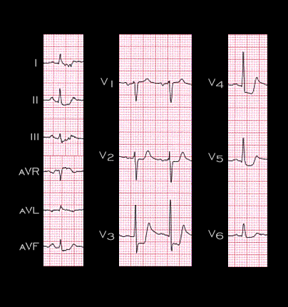
This ECG was shown on the first two pages of this section (6.1.0 and 6.1.1). It demonstrates depression of the ST segment in leads I, II, III, aVF and in leads V2-V6, reflecting non-transmural ischemia. The changes were brought on by exertion in a patient with a high grade lesion in the circumflex coronary artery. The diffuseness of ST segment depression is an indication of the inability of these changes to localize the ischemic region or the vessel involved. It should be noted however, that in patients with chest pain occurring at rest, diffuse ST segment depression, especially when it is coupled with ST elevation in lead aVR, as in this example, may indicate severe 3 vessel disease or sub-total occlusion of the left main coronary artery.

Book traversal links for 6.1.47 (46)

  • 6.1.46 (45)
  • Up
  • 6.1.48 (47)

Site is under construction

Book navigation

  • Introduction to First Edition
  • 1.0.0 Generation of the ECG
  • 2.0.0 The Normal Electrocardiogram
  • 3.0.0 Inter and intra-ventricular Conduction Disturbances
  • 4.0.0 Ventricular Hypertrophy
  • 5.0.0 ELECTROLYTE ABNORMALITIES, DRUG EFFECTS AND THE LONG QT SYNDROMES
  • 6.0.0 Ischemia and Infarction - Introduction (frame i and ii)
    • 6.1.0 ST Segment (frame1)
      • 6.1.1 (2)
      • 6.1.2(3)
      • 6.1.3(4)
      • 6.1.4(5)
      • 6.1.5(6)
      • 6.1.6 (7)
      • 6.1.7 (8)
      • 6.1.8 (new frame)
      • 6.1.9 (9)
      • 6.1.10 (frame 10)
      • 6.1.20 (frame 20)
      • 6.1.30 (frame 30)
      • 6.1.40 (frame 39)
        • 6.1.41(frame 40)
        • 6.1.42 (41)
        • 6.1.43 (42)
        • 6.1.44 (43)
        • 6.1.45 (44)
        • 6.1.46 (45)
        • 6.1.47 (46)
        • 6.1.48 (47)
        • 6.1.49 (48)
      • 6.1.50 (49)
    • 6.2.0 Q WAVES (frame 53)
    • 6.3.0 T WAVES (frame 75)
    • 6.4.0 (Bundle Branch Blocks and othe IVCDs--frame 87)
    • 6.5.0 (frame 106) FALSE POSITIVES
  • 7.0.0 Tachycardias- Introduction
  • 8.0.0 The Bradycardias frame i-introduction
  • 9.0.0 The ECG of Heart Murmurs-introduction
  • 10.0.0 The Electrocardiogram in the Emergency Department-Introduction
Powered by Drupal