Home
Understanding the Electrocardiogram

Main navigation

  • Home
  • Understanding ECGs
    • Introduction
User account menu
  • Log in

Breadcrumb

  1. Home
  2. Understanding the Electrocardiogram In Health & Disease
  3. 6.0.0 Ischemia and Infarction - Introduction (frame i and ii)
  4. 6.1.0 ST Segment (frame1)
  5. 6.1.40 (frame 39)

6.1.42 (41)

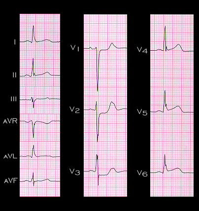
Involvement of the posterior wall may also accompany lateral wall ischemia and infarction. The ECG shown here is from a 44 year old male with chest pain. There is slight ST elevation in leads I and aVL and more obvious ST elevation in leads V5 and V6. This indicates acute transmural ischemia/infarction of the lateral wall. There is also marked ST depression in leads V2 and V3 and lesser ST depression in V1. These changes are the reciprocal of the ST elevation the would be expected in leads placed on the left posterior chest wall, opposite leads V2 and V3, i.e. posterior leads placed in the V8 and V9 positions, and indicate posterior wall involvement. Note that there are no ST segment changes in leads II, III or aVF, the inferior leads. This observation suggests that blood flow the in the posterior descending coronary artery was preserved and that the culprit lesion was not in its parent vessel, i.e, either the right or circumflex coronary artery.

Book traversal links for 6.1.42 (41)

  • 6.1.41(frame 40)
  • Up
  • 6.1.43 (42)

Site is under construction

Book navigation

  • Introduction to First Edition
  • 1.0.0 Generation of the ECG
  • 2.0.0 The Normal Electrocardiogram
  • 3.0.0 Inter and intra-ventricular Conduction Disturbances
  • 4.0.0 Ventricular Hypertrophy
  • 5.0.0 ELECTROLYTE ABNORMALITIES, DRUG EFFECTS AND THE LONG QT SYNDROMES
  • 6.0.0 Ischemia and Infarction - Introduction (frame i and ii)
    • 6.1.0 ST Segment (frame1)
      • 6.1.1 (2)
      • 6.1.2(3)
      • 6.1.3(4)
      • 6.1.4(5)
      • 6.1.5(6)
      • 6.1.6 (7)
      • 6.1.7 (8)
      • 6.1.8 (new frame)
      • 6.1.9 (9)
      • 6.1.10 (frame 10)
      • 6.1.20 (frame 20)
      • 6.1.30 (frame 30)
      • 6.1.40 (frame 39)
        • 6.1.41(frame 40)
        • 6.1.42 (41)
        • 6.1.43 (42)
        • 6.1.44 (43)
        • 6.1.45 (44)
        • 6.1.46 (45)
        • 6.1.47 (46)
        • 6.1.48 (47)
        • 6.1.49 (48)
      • 6.1.50 (49)
    • 6.2.0 Q WAVES (frame 53)
    • 6.3.0 T WAVES (frame 75)
    • 6.4.0 (Bundle Branch Blocks and othe IVCDs--frame 87)
    • 6.5.0 (frame 106) FALSE POSITIVES
  • 7.0.0 Tachycardias- Introduction
  • 8.0.0 The Bradycardias frame i-introduction
  • 9.0.0 The ECG of Heart Murmurs-introduction
  • 10.0.0 The Electrocardiogram in the Emergency Department-Introduction
Powered by Drupal