
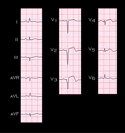
This ECG was recorded 3 weeks later from the same patient. It shows persistent ST segment elevation in leads V1-V4 and inverted T weaves in leads V3 and V4. A QS complex is now present in lead V3 as well as in leads V1 and V2 and a small Q wave is present in V4. These changes are consistent with an infarction of the left ventricular wall within the distribution of the left anterior descending coronary artery. The persistence of ST segment elevation 3 weeks after the acute event suggests the possibility of a ventricular aneurysm.