Home
Understanding the Electrocardiogram

Main navigation

  • Home
  • Understanding ECGs
    • Introduction
User account menu
  • Log in

Breadcrumb

  1. Home
  2. Understanding the Electrocardiogram In Health & Disease
  3. 2.0.0 The Normal Electrocardiogram
  4. 2.2.0 PR Interval

2.2.10

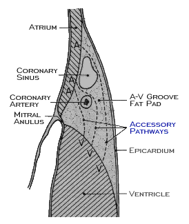
Conduction across the AV junction may also be accelerated, causing shortening of the PR interval to less than 120 ms. This may be due to accelerated conduction within the AV node itself,  considered to represent one end of the normal physiologic spectrum,  or to conduction across the AV junction via a tract or tracts of myocardial tissue, known as Kent bundles that bypass the AV node, These AV nodal bypass tracts are also referred to as accessory pathways and may span the AV ring at a variety of locations. Impulses conducted to the ventricles via these pathways activate the ventricles abnormally and cause intra-ventricular conduction disturbances with characteristic ECG features. These are discussed in greater detail in the next chapter.

Book traversal links for 2.2.10

  • 2.2.9
  • Up
  • 2.3.0 QRS Complex

Site is under construction

Book navigation

  • Introduction to First Edition
  • 1.0.0 Generation of the ECG
  • 2.0.0 The Normal Electrocardiogram
    • 2.1.0 P Wave
    • 2.2.0 PR Interval
      • 2.2.1
      • 2.2.2
      • 2.2.3
      • 2.2.4
      • 2.2.5
      • 2.2.6
      • 2.2.7
      • 2.2.8
      • 2.2.9
      • 2.2.10
    • 2.3.0 QRS Complex
    • 2.4.0 ST segment
    • 2.5.0 T wave
    • 2.6.0 QT Interval
    • 2.7.0 U Wave
  • 3.0.0 Inter and intra-ventricular Conduction Disturbances
  • 4.0.0 Ventricular Hypertrophy
  • 5.0.0 ELECTROLYTE ABNORMALITIES, DRUG EFFECTS AND THE LONG QT SYNDROMES
  • 6.0.0 Ischemia and Infarction - Introduction (frame i and ii)
  • 7.0.0 Tachycardias- Introduction
  • 8.0.0 The Bradycardias frame i-introduction
  • 9.0.0 The ECG of Heart Murmurs-introduction
  • 10.0.0 The Electrocardiogram in the Emergency Department-Introduction
Powered by Drupal