Home
Understanding the Electrocardiogram

Main navigation

  • Home
  • Understanding ECGs
    • Introduction
User account menu
  • Log in

Breadcrumb

  1. Home
  2. Understanding the Electrocardiogram In Health & Disease
  3. 8.0.0 The Bradycardias frame i-introduction
  4. 8.2.0 SA Block (frame 41)

8.2.1 (42)

Electrodes placed directly over the sinus node in experimental animals and in patients undergoing cardiac surgery have been used to record the electrical signals associated with sinus node depolarization. These studies have shown that the normal interval from sinus node depolarization to the onset of the P wave is from 25 to 50 ms. (Hariman el al, Circulation 61:1024,1988).

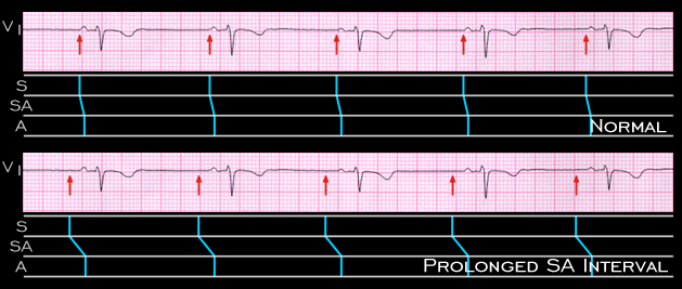
In the tracing from the 23 year old female, shown again here, the atrial rate is 44 and there is 1:1 AV conduction with a normal PR interval (0.18 seconds). The relationship of the sinus node impulse (arrows) to the onset of the P wave, the SA interval, may be normal, as depicted in the upper ladder diagram. However, it is impossible to exclude the possibility that the SA interval is prolonged (i.e.1st degree SA block), as depicted in the lower diagram. A third possibility is shown on the next page.

Book traversal links for 8.2.1 (42)

  • 8.2.0 SA Block (frame 41)
  • Up
  • 8.2.2 (43)

Site is under construction

Book navigation

  • Introduction to First Edition
  • 1.0.0 Generation of the ECG
  • 2.0.0 The Normal Electrocardiogram
  • 3.0.0 Inter and intra-ventricular Conduction Disturbances
  • 4.0.0 Ventricular Hypertrophy
  • 5.0.0 ELECTROLYTE ABNORMALITIES, DRUG EFFECTS AND THE LONG QT SYNDROMES
  • 6.0.0 Ischemia and Infarction - Introduction (frame i and ii)
  • 7.0.0 Tachycardias- Introduction
  • 8.0.0 The Bradycardias frame i-introduction
    • 8.1.0 Sinus Bradycardia and AV block (frame 1)
    • 8.2.0 SA Block (frame 41)
      • 8.2.1 (42)
      • 8.2.2 (43)
      • 8.2.3(44)
      • 8.2.4 (45)
      • 8.2.5 (46)
      • 8.2.6 (47)
      • 8.2.7 (48)
      • 8.2.8 (49)
      • 8.2.9 (50)
      • 8.2.10 (frame 51)
      • 8.2.11 (52)
      • 8.2.12 (53)
      • 8.2.13 (54)
      • 8.2.14 (55)
      • 8.2.15 (56)
    • 8.3.0 Group Beating (frame 57)
  • 9.0.0 The ECG of Heart Murmurs-introduction
  • 10.0.0 The Electrocardiogram in the Emergency Department-Introduction
Powered by Drupal